– a possible alternative to surgically implanted surface electrodes?
Wouldn’t it be great if we could reliably measure our brain’s activities without the need to open the skull? An Australian team is exploring this possibility by adapting a technology from cardiology: A stent, inserted through a catheter into a cerebral blood vessel in the brain. The team modified the stent so that it could function as an electrical recording electrode with various, independent channels.
But how good can such endovascular recordings get? Could they one day replace more conventional recording techniques that are used nowadays, e.g., in preparation for a brain surgery, or for brain-machine interfaces?
To answer this question, the team performed a systematic comparison of the signal qualities recorded from their “stentrodesTM“ and those from more conventional flat electrode arrays that were implanted onto the surface of the brain, either epidurally or subdurally. In this comparison, it is fair to assume that the spatial characteristics of the electrodes matter. So, CorTec custom-designed flat electrode arrays for this purpose that had exactly the same or only slightly larger or smaller diameters as the endovascular electrodes.
The electrode type that tended to perform best in the authors’ comparison (esp. signal to-noise-ratio) was the subdural type. That does not come as a big surprise, since it is closest to the electrically active neurons and there is no dura or blood vessel wall separating it from the neural tissue. What was surprising, however, was that the differences between all electrode types were quite small and, actually, not statistically significant. The study concludes that the exact placement of electrodes, be it subdurally, epidurally, or even endovascularly, does not matter very much for signal quality.
Therefore, the authors argue that for medical use, especially for implanted neuromodulation devices, one may consider the least invasive options for placing recording electrodes, including their novel endovascular electrodes that do not require any brain surgery.
Citation:
John SE, Opie NL, Wong YT, Rind GS, Ronayne SM, Gerboni G, Bauquier SH, O’Brien TJ, May CN, Grayden DB, Oxley TJ. (2018): Signal quality of simultaneously recorded endovascular, subdural and epidural signals are comparable. Sci Rep. 2018 May 30;8(1):8427. doi: 10.1038/s41598-018-26457-7. Erratum in: Sci Rep. 2018 Nov 27;8(1):17469.
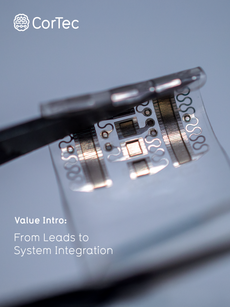
Learn more about possible applications with our °AirRay Grid Electrodes